
Photon-counting CT Scanner: Siemens NAEOTOM Alpha
Dual Source Photon Counting Detector Computed Tomography (PDCT) Scanner is located in MRF54.

Xenon-129 Hyperpolarizer: Polarean Model 9820
This is a dedicated system for the purpose of polarization of Xenon used for the assessment of regional lung structure and function via magnetic resonance imaging (MRI). The polarizer is located in L543 PBDB.

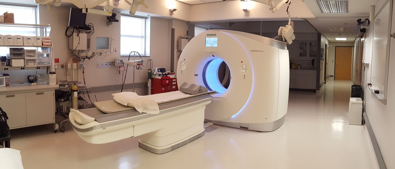
Procedure Room
The Procedure Room is located in 70 MRF.

DEXA scanner: Hologic Horizon A
The research-dedicated dual-energy X-ray absorptiometry (DEXA) scanner: Hologic Horizon A is located in 158 MRF.

C-arm Fluoroscope: OEC 990 Elite
C-arm Fluoroscope: OEC 990 Elite is located in 60 MRF.
Quantitative Software
Pulmonary Analysis Software Suite (PASS)
Our Pulmonary Analysis Software Suite has been developed by in house programmers. It is a comprehensive package for the manipulation, display and analysis of multidimensional image data sets. PASS is intended to assist doctors and researchers in the field of pulmonary research with lung segmentation, histogram analysis, low attenuation area measurement, nodules and lung tissue classification, time sequence image analysis (TSIA), phantom analysis, modulation transfer function (MTF) and noise-power spectrum (NPS) analysis.
VIDA Insights
VIDA Insights is a commercial, FDA approved software for quantitative lung analysis (VIDA Insights – Clinical Imaging Intelligence). We have expert analysts capable of processing CT data with VIDA Insights and providing quality control. Metrics available can capture lung density, airways, gas trapping, mucus plugging.
Siemens Recon CT
In typical CT scanning, reconstruction parameters are selected during data acquisition and then reconstructed data in DICOM format is transferred to the user. Raw data is not routinely saved or transferred. However, if additional alternate reconstructions may be needed post-acquisition, raw CT data should be stored, such that the research dedicated Siemens Recon CT can be used to generate additional alternate reconstructions of the data without requiring time on the CT scanner system.
PVS
Procedural Verification Software - PVS - is an automated web portal system for tracking and verifying scan parameter data for multi-center studies. A study participant is entered into PVS which in turn provides study and site specific information, including scanner and scan parameters to be used. A printout of this information is printed and provided to the study certified CT technologist. Once the scan is completed, the post scan information (including FOV, recon kernel, effective dose, etc.) is logged and the information is transmitted to the study's Radiology Center. PVS users can register subjects for their site in the database, communicate with the Radiology Reading Center via messaging system, and view details of previous scans performed by their site, aiding in the assurance of longitudinal continuity.
MIFAR
Medical Imaging File Archive Recovery - MIFAR - is a set of software tools for storing medical image data along with other associated post-image processing files, physiologic records, and lab notes.