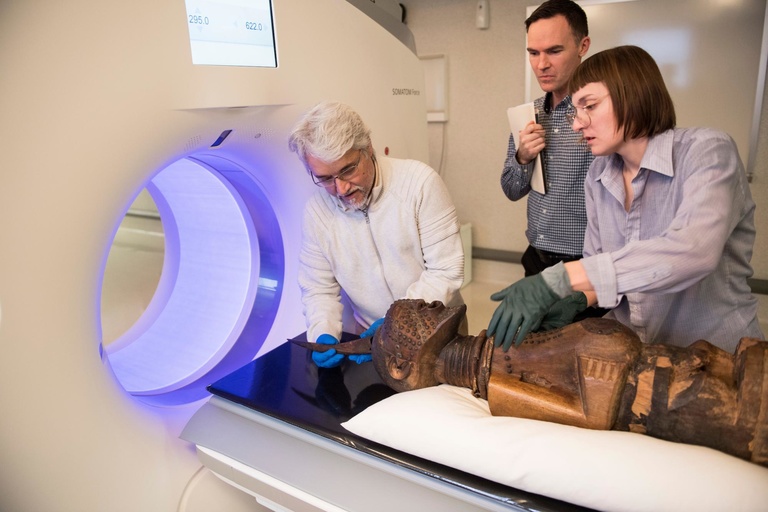
Thanks to imaging technology, museum staff at the University of Iowa Stanley Museum of Art have a better idea of what might be inside 16 of the museum's works of African art—without having to break them open.
The works, which were analyzed by a computed tomography (CT) scanner in January at the University of Iowa Hospitals & Clinics, are power objects that date from the late 19th century to mid-20th century and are made from materials such as wood, clay, metal, horn, and animal skin.
However, that's just what can be seen by the naked eye. What's hidden inside the objects truly gives them their importance. The sculptures contain cavities into which substances empowered by a diviner were placed.
The scans were done on the Siemens Somatom Force CT scanner, which was installed in the University of Iowa Hospitals & Clinics' Advanced Pulmonary Physiomic Imaging Laboratory (APPIL) in 2015. It's a dual-energy scanner, which means it can scan and produce images of an object at two energy levels.
"That allows us to visualize different materials based on those energy levels," says Melissa Saylor, a CT research associate in the lab. "It's great for something like this because we didn't know what types of materials were inside."
The scanner also uses metal artifact reduction, which reduces streaking that can occur from metal objects and allows for better detail. This was beneficial as some of the art contained metal pieces, such as nails.
UI Stanley Museum of Art staff use health care technology to learn more about the contents and construction of 'power objects'
Sunday, October 29, 2023


